Getting a Clearer Picture of Deep Sea Samples: New Lab Imaging Equipment Gives a Closer Look at E/V Nautilus Deep Sea Specimens

This blog was written by NA181 Lead Science Communication Fellow Erin Winick Anthony.
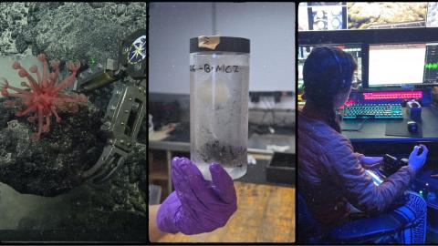
To keep up with the latest scientific developments, E/V Nautilus must consistently upgrade the technology on board. Sometimes that’s the large equipment, like the new multibeam echosounder installed during the off-season this past year, which has enabled wider and deeper mapping throughout the 2026 season. Other times, it’s the smaller upgrades, like the ones seen in the wet lab for the first time during NA181. The lab received a new microscope and photography setup to analyze and capture images of samples taken from the deep sea.
During this expedition, science team members Paula Rodriguez Flores and Jessica Eberle implemented the new imaging hardware, so we sat down and asked them a few questions about the setup.
Check out a photo album of some of the microscopic photos from the expedition here.
What is the change in equipment? What microscope and photography setup did we have before, and what do we have now?
Jessica: Before setting up this camera and stereoscope system, we mainly used an iPad for sample imaging. The iPad was a quick, easy way to image many specimens, but our new setup greatly improves image quality. We now have a Canon EOS RP Mirrorless camera body, Leica EZ4W microscope, standard and macro lenses, flashers, flash trigger, and more.
This is a massive improvement in our onboard imaging capabilities. Now we can image anything from teeny tiny 1mm coral polyps to "Bertha", an enormous rock sample from the expedition. The flip side of this more technical setup is that dialing in the lighting and camera settings takes extra time for both initial testing and imaging. But the extra effort is worth it; everyone using these photos later will get much higher-quality images.
In general, why are microscopy and photography important for the scientific work we do on board?
Paula: Chromatophore pigments, particularly in shrimps, squat lobsters, hermit crabs, and many peracarids (such as isopods, amphipods, and mysids), dissolve in ethanol, causing specimens to lose their coloration and turn white after fixation. Because color can be an important taxonomic character, it is therefore essential to photograph these crustaceans while they are still alive or freshly collected, using macrophotography.
A clear general photograph showing the animal's overall appearance, including the carapace and legs, is a valuable addition to a taxonomic description. Detailed photographs of specific taxonomic characters, along with their fresh coloration, provide additional information and are an important component of high-quality species descriptions. This is particularly valuable not only for describing new species, but also for previously described species for which fresh coloration and morphological details are still unknown, as is often the case with deep-sea species.
Why is this equipment upgrade helpful for science teams on board?
Jessica: It is essential to our work. High-quality images before preservation increase the scientific value of every specimen, especially the biological ones. It preserves a record of their living morphology and color that can never be replicated once they leave the deck. We want to do the highest-quality science possible during our time at sea, so tools that improve data collection are invaluable. These images also remain permanently linked to the specimens, so researchers can access them long after the samples leave the ship and are processed in other labs. The photos can also identify samples if a mix-up happens, so they're an important record all around.
Paula: I think it is very important to facilitate and support research on deep-sea organisms as science and research methods continue to evolve to address new questions. Scientific methods are not static, and this is particularly true for deep-sea animals. Every day, we are developing new approaches to preserve specimens and new ways to study their morphology, genetics, and genomics.
I think it is great that the ship continues to upgrade its capabilities, making the science more inclusive and keeping shipboard research up to date with the latest techniques and approaches used by scientists on shore. I am very happy and grateful to have witnessed this upgrade, and I am really excited to see the results and the new scientific opportunities it will open up.
Do you have any examples of how using the new equipment was helpful on this expedition?
Jessica: Paula was super excited to use the setup for her squat lobsters, which also lose their pigmentation in ethanol. Because color can be a key diagnostic trait for species identification, this setup allowed us to take publication-worthy images while their colors were still vibrant - images she will use for species descriptions. We also photographed the squat lobsters in situ alongside their host corals, documenting ecological associations we discovered only through visual exploration of these deep-sea environments.
The new stereoscope also served as a useful tool for imaging and identifying crystal inclusions in rock samples! It also allowed us to take close-ups at up to 35x magnification of important morphological features like coral sclerites and squat lobster spines.
Paula: It has helped enormously. We have been able to obtain not only high-resolution images of the general appearance and morphology of the specimens, but also detailed photographs of important taxonomic characters while the animals are still fresh. This provides information that would otherwise be impossible to observe once the specimens are preserved in scientific collections.
For example, we can document highly intriguing pigmentation patterns in the crustacean eyes, as well as pigments present on specific body segments. This information is valuable not only for taxonomic studies but also for understanding the evolution and ecology of these deep-sea organisms. This upgrade in the photographic camera system provides a unique opportunity to create a permanent visual record of specimens and their details. It will also help attract other taxonomists to study and work on this valuable material.
Why is it important to keep upgrading and improving our capabilities on the ship?
Paula: I think it is important to support research on deep-sea organisms as science and research methods continue to evolve to address new questions. Scientific methods are not static, and this is particularly true for deep-sea animals. Every day, we are developing new approaches to preserve specimens and new ways to study their morphology, genetics, and genomics.
I think it is great that the ship continues to upgrade its capabilities, making the science more inclusive and keeping shipboard research up to date with the latest techniques and approaches used by scientists on shore. I am very happy and grateful to have witnessed this upgrade, and I am really excited to see the results and the new scientific opportunities it will open up.
What future improvements do you hope to see in scientific equipment on board the ship?
Jessica: For molecular and eDNA work, adding a Milli-Q system to have true ultra-pure water would be a game-changer, alongside updates to the shipboard saltwater system (it's a "wee bit" rusty). I'm also a big fan of "underway" seawater sensors that monitor temperature, salinity, O2, Chla, etc., to passively collect environmental data as we go.
For the new photography setup, I would love to add deeper or taller, clear-sided imaging containers so larger specimens stay fully submerged without shading, along with light diffusers to soften the light. We're still learning as we go, but small things like this would further improve the setup and help us produce the best, most consistent scientific data possible.
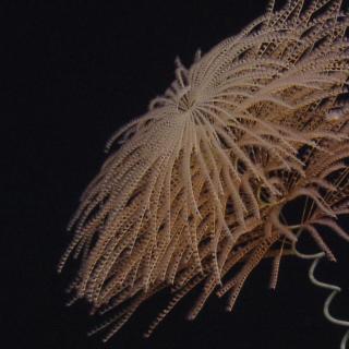
Exploration of Wake Island's Deep Sea
Located roughly midway between the Hawaiian and Mariana Islands, Wake Island is one of the most isolated land masses on Earth. While there have been some recent expeditions to this remote region, the vast majority of the 407,241 square kilometers of seafloor surrounding Wake remains unmapped and uncharacterized, thereby representing one of the most poorly surveyed areas under US jurisdiction.